

Permanent Pacemaker Implantation
Abnormally slow heart rates and rhythms may require permanent pacemaking, using one, two or three wires, or leads. These are placed into the heart using veins which run from the shoulder region below the collarbone.
Permanent Pacemaking has been available for abnormally slow heart rhythms for over 50 years and has been greatly refined over that time.
Dual-chamber permanent pacemaker
Single-chamber permanent pacemaker
Abnormally slow heart rhythms which may require pacing include disorders of conduction such as 3rd Degree Heart Block or disorders of impulse formation such as Sinus Node dysfunction. The ultimate decision to implant a pacemaker requires a complete review of each patient’s specific case by the Electrophysiologist.
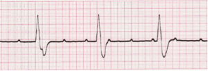
Rhythm strip showing 3rd Degree Heart Block. Note the lack of co-ordination between the atrial and ventricular signals.

Rhythm strip showing a pause in the heart rhythm.
By far the most common form of permanent pacemaking involves placing the pacing wires (or leads) through a large vein just below the collarbone, to the heart. This is done by making a small incision just below the collarbone to access this vein, then using a form of continuous X-ray camera called a fluoroscope to guide the wires into position.
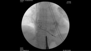
Fluoroscopic image showing pacemaker leads in place in the heart.
The Permanent Pacemaker itself is placed below the skin surface after connection to the pacing wires and before the incision is closed. In the end, only a small scar is visible, although the pacemaker may be felt below the skin in thinner patients.

The surgery itself last approximately one hour and is a same-day admission and discharge in most cases. Deep conscious sedation and local anaesthetic are used to maximize patient comfort. Pacemaker implantation is performed in the Electrophysiology Suite as well as the general Operating Room.
A paced heart rhythm does not feel any different to a non-paced rhythm, but an Electrocardiogram will show the pacemaker’s activity.

Rhythm strip showing pacemaker activity in both the upper and lower chambers of the heart
Permanent pacemakers may use one, two or even three leads depending on the heart rhythm abnormality and the strength of the patient’s heart muscle itself.

Chest X-ray showing dual-chamber pacemaker
Patient who have permanent pacemakers are followed up in the Cardiac Device Clinic, typically every 6 or 12 months. Cardiac Device Clinics are available at both the Credit Valley and the Mississauga Hospital Site at Trillium Health Partners.

